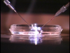
Many couples who can’t manage to conceive by themselves often turn to fertility clinics to help them to have a child. One of the most common ways that fertility clinics help is by performing in vitro fertilization (IVF). Since the first baby born by IVF – Louise Brown – came into the world in 1978, there have been enormous advances in the field, but the technique still isn’t always successful. In fact, a study done in the UK showed that only about 24% of women who have an IVF embryo implanted actually give birth to a live baby. This type of rate is not specific to the UK – if you are looking for a reproductive endocrinologist Kansas City success rates are likely to be quite similar.
However, it looks like there may be some new hope on the horizon – and again, it is coming from the UK. The use of IVF is very widespread there, with about 60,000 couples having the treatment each year. Because of this, there is a lot of research carried out in the UK on IVF, including at a number of UK fertility clinics. One of those, CARE Fertility Group, now seems to have made a breakthrough that may raise the success rate for IVF to as high as 78%.
The technique is relatively simple. When an egg is fertilized using IVF, it is left for a period of time to develop into an early-stage embryo before it is implanted into the womb. Normally, as long as the embryo appears to be viable at the end of this waiting period, it is implanted without any other checks. With the new technique, the fertility clinic takes thousands of time-lapse pictures of the embryo during the first few days of its life, and uses the results to determine whether or not the embryo is likely to develop successfully when it is implanted.
When the fertility clinic first starts to take these photographs, the embryo only consists of a few cells. However, it rapidly develops into a fluid-filled ball, known as a blastula. A little later on, this blastula starts to become a more complicated structure known as a gastrula, which consists of three layers – instead of the one found in the blastula. These layers grow to form different parts of the embryo. The outer ectoderm develops into the nervous system, as well as other body structures such as the lining of the mouth and the skin. The middle layer, the mesoderm, gives rise to muscles as well as parts of the genitalia. The inner layer, the endoderm, forms the gut and the lungs, as well as many of the body’s internal organs – such as the liver and pancreas.
It turns out that the amounts of time between the blastula first forming and the embryo emerging is a good indicator of whether or not implanting the embryo will be successful. Normal embryos take less than six hours to do this – and this is really the basis of the breakthrough. If an embryo takes longer than six hours, then it is likely to develop a condition called aneuploidy, which is when there are an abnormal number of chromosomes in the embryo’s cells. It is this chromosome count that is associated with much higher pregnancy failure rates. In fact, abnormal numbers of chromosomes are also found in other medical conditions – for example, Down’s syndrome is a result of having three copies of chromosome 21, rather than the normal two copies.
By determining how long this gap was in embryos that were waiting to be implanted, the fertility clinic researchers were able to raise the success rate in the group that they studied all the way up to 78%. This confirms the results of a previous study that looked retrospectively at a group of 69 women who had embryos implanted – this concluded at that time that 61% of them could have been helped by measuring the gap between the two points in their embryo’s development.
According to Professor Simon Fishel, who heads up the fertility clinic where the technique was developed, “Our work has shown that we can easily classify embryos into low or high risk of being chromosomally abnormal. This is important because in itself this is the largest single cause of IVF failure and miscarriage. The beauty of this technology is that the information is provided by a non-invasive process. So far we have seen a 56 per cent uplift compared to conventional technology, giving our patients the equivalent to a 78 per cent live-birth rate.”
Originally posted on January 16, 2014 @ 7:12 pm

